The new Opto Magis Hardware is much more robust and has greater processing capacity.
The processor used is a 1.8 GHz Quadcore CPU is a Quad-Core RAM: DR3 It has two interfaces, one HDMI and one Micro USB Resolution of 1,920 x 1,080 Works with Android 4.4.2 operating system
NEW MAGIS
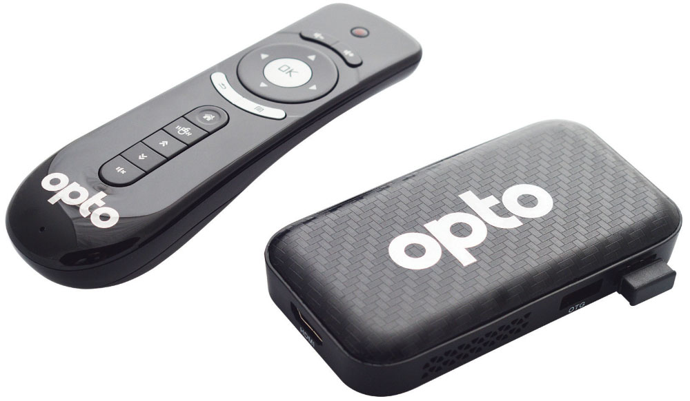
1) Opto Magis mini computer.
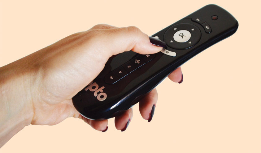
2) Wireless remote control.
3) Wireless receiver.
4) AAA batteries (02 units).
5) Power supply with USB thermal 6. HDMI cable.
7) Blue/Red 3D Glasses.
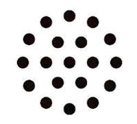
Tables
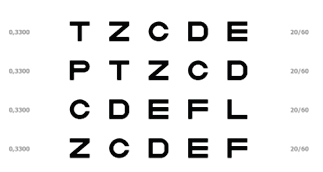
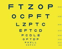
Letters
This is the most commonly used table for visual acuity exams.
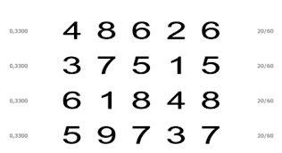
Numbers
This is the same type of letter chart, but with numbers. There is no difference in performing the exam using the letter chart or the number chart.
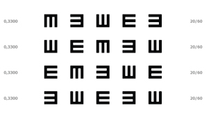
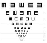
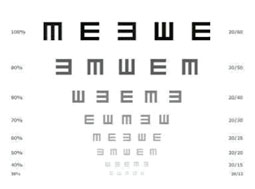
Snellen E Chart
The E’s table is used to measure the visual acuity of illiterate adults when tables with symbols or drawings are not required.
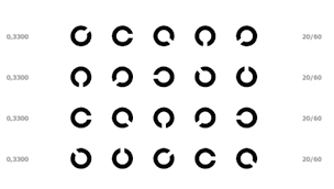
Table with Landolt’s C
The Landolt C chart was created by the Swiss ophthalmologist Edmund Landolt. It is also an optotype chart for illiterate adults or children. It consists of “broken” circles in different positions. Studies indicate that there is very little difference between measuring visual acuity with the Snellen E, Letter, Number charts and the Landolt “C” chart.
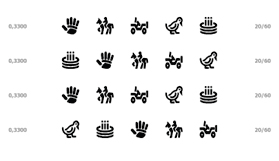
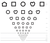
Allen Symbol Tables and Optotypes
These are tables for illiterate children.
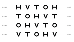
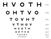
HVTO Table
This is another optional chart for non-literate children. It is a chart widely used for screening.
Functions
Functions are masks and operations that modify the tables allowing the examiner to improve the measurement of the patient’s visual acuity.
Função Longe/Perto (Far-Near)
Durante a instalação do Opto Magis deve-se configurar duas distâncias bases para trabalho (ele já vem de fábrica com as distâncias 6.0 metros para longe e 1.5 metros para perto que podem ser modificadas).
A função Longe/Perto (Far-Near) irá ajustar as dimensões das máscaras para essas duas distâncias pré-programadas. A Função Longe é utilizada para testes em pessoas normais e a Função Perto para pessoas com visão subnormal.
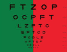
Green-Red Mask
The Red-Green (also called the Duo-Chrome test) is a color mask applied to tables. It can be applied to all tables.
This test is intended to check whether the client is hyper or hypo refracted during the refraction exam.
The principle of this test is based on the fact that green light refracts more and red light refracts less.
If a patient with myopia sees the optotypes sharper on the green side than on the red side, this means that the eye is hyperrefracted. For example, if it needs -2.00 and is at -2.50, it will see the letters on the green frame better. Then it must be corrected until the patient sees both sides with approximately the same clarity.
In hyperopia , the verification is the opposite of that of myopes. When the patient sees the letters better on the green side, he is hypo refracted and when he sees better on the red side, he is hyper refracted.
Green-Red is especially useful when the patient appears indecisive or tired of the tests or the examiner himself feels insecure.
The case of myopic people is a good example because the myopic person accommodates a lot and makes correct refraction difficult and red-green will help to clear up doubts.
ETDRS Standard
ETDRS is a standard for presenting visual acuity charts.
In this pattern, the optotypes are presented in lines whose size decreases from one line to the next.
In this pattern, the distances between optotypes that are in the same line and the distance between the lines are proportional to the size of the optotypes.
This type of table presentation is widely used in controlled clinical studies.
In Opto Magis, all tables can be presented in this standard, including combined with the Green-Red test.
Blue-Yellow Mask
In this mask, which can be applied to all optotype tests, the optotypes are presented in blue on a yellow background. Diseases related to the aging of the lens make it difficult to see in Blue/Yellow mode. The test can also be used to distinguish diseases such as Optic Neuritis (inflammation of the optic nerve) from Papilledema (swelling of the papilla). Papilledema. In papilledema, Blue/Yellow vision is difficult. In Optic Neuritis, it is not.
Contract Sensitivity
This is not exactly a mask, but an aspect that can be applied to all optotype tests.
In this test, the optotypes are presented at different intensities (different contrasts in relation to the background) adjusted by the examiner.
The contrast sensitivity test complements the conventional visual acuity test, allowing a more accurate assessment of the patient’s visual condition before and after surgeries such as cataract and refractive surgeries, helping the examiner to better guide the patient regarding their expectations.
This test is not very accurate when performed on people without contrast sensitivity problems, but it decisively indicates patients with low sensitivity.
Tests
The tests are figures that allow the examiner to perform complex examinations that help improve the performance of the visual acuity examination and determine the existence of diseases and vision defects.
Astigmatism Test 1
This test will identify whether the patient has astigmatism, that is, does not see a group of lines clearly. The examiner should ask the patient about which lines he sees clearly and which he sees “fuzzy” (out of focus and/or not sharp). The image on the side shows how the patient will see the lines in a fuzzy way, indicating that he has astigmatism.
Astigmatism Test 2
Same test as above with simple lines.
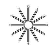
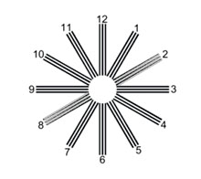
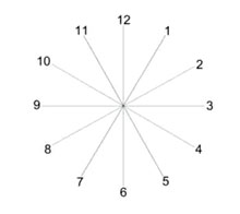
Torsion Test
Motor test to measure and classify a cyclotorsion (ocular deviation where the angle deviates in its anteroposterior axis, with incyclodeviation, the vertical meridian of the cornea deviating inwards, and in excyclodeviation, outwards). The patient must use three-dimensional vision glasses to perform this test.
In the view of a patient with cyclotorsion, the bars are not parallel. The examiner will tilt the bars until the patient sees them parallel. The tilt of the bars will indicate the level of cyclotorsion. This examination is done for each eye separately.
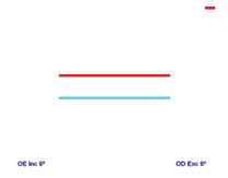
Cross Cylinder
Test used to determine and/or refine the axis and power of the patient’s astigmatism. In this test, the examiner asks the patient which of the two cylinder options presented (lenses with cylinder placed on the greens) the circles are more round or less oval.”
Stereopsis Test
In this test, the evaluator will assess the patient’s three-dimensional perception.
This is a very important test for evaluating operators of machines such as forklifts, driving machines, etc.
Using the glasses and for each figure, the examiner will start the test with the figures as separate as possible and will join them together. When the patient indicates that for him the shapes are already together, the exam will indicate the patient’s degree of stereopsis.
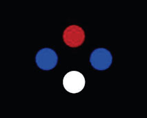
Worth Lights Test
Worth’s four-light test is a clinical test for left or right eye suppression. Suppression occurs during binocular vision when the brain cannot process the information received from one eye. In other words, this test determines the presence of binocular vision.
It consists of four illuminated circles (two blue, one red and one white) on a black background. The test is performed binocularly, using 3D glasses. The following cases may occur during the test:
1° When the observer sees two circles (red and white), he lacks vision with the eye with the blue lens.
2° When the observer sees three circles (the blue ones and the white one), he lacks the vision of the eye with the red filter.
3° When the observer sees four circles, he has binocular vision.
4° When the observer sees five circles, there is the presence of diplopia.
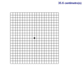
Amsler grid
Simple and quick method for evaluating the macular visual field. The patient will see gaps in the grid or distorted areas.
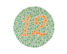
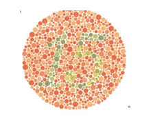
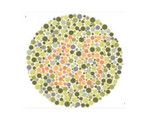
Ishihara Card Test
The Ishihara Color Test is a test for detecting color blindness. The test consists of showing a series of colored cards, each containing several circles made of colors slightly different from those located nearby.
Depending on the patient’s response, the system indicates whether there are color blindness problems.
Utilizamos ferramentas e serviços de terceiros que utilizam cookies. Essas ferramentas nos ajudam a oferecer uma melhor experiência de navegação no site. Ao clicar no botão “Aceitar” ou continuar a visualizar nosso site, você concorda com o uso de cookies em nosso site.
This website uses cookies to improve your experience while you navigate through the website. Out of these, the cookies that are categorized as necessary are stored on your browser as they are essential for the working of basic functionalities of the website. We also use third-party cookies that help us analyze and understand how you use this website. These cookies will be stored in your browser only with your consent. You also have the option to opt-out of these cookies. But opting out of some of these cookies may affect your browsing experience.
Necessary cookies are absolutely essential for the website to function properly. These cookies ensure basic functionalities and security features of the website, anonymously.
Cookie
Duração
Descrição
cookielawinfo-checbox-analytics
11 months
This cookie is set by GDPR Cookie Consent plugin. The cookie is used to store the user consent for the cookies in the category "Analytics".
cookielawinfo-checbox-functional
11 months
The cookie is set by GDPR cookie consent to record the user consent for the cookies in the category "Functional".
cookielawinfo-checbox-others
11 months
This cookie is set by GDPR Cookie Consent plugin. The cookie is used to store the user consent for the cookies in the category "Other.
cookielawinfo-checkbox-necessary
11 months
This cookie is set by GDPR Cookie Consent plugin. The cookies is used to store the user consent for the cookies in the category "Necessary".
cookielawinfo-checkbox-performance
11 months
This cookie is set by GDPR Cookie Consent plugin. The cookie is used to store the user consent for the cookies in the category "Performance".
viewed_cookie_policy
11 months
The cookie is set by the GDPR Cookie Consent plugin and is used to store whether or not user has consented to the use of cookies. It does not store any personal data.
Functional cookies help to perform certain functionalities like sharing the content of the website on social media platforms, collect feedbacks, and other third-party features.
Performance cookies are used to understand and analyze the key performance indexes of the website which helps in delivering a better user experience for the visitors.
Analytical cookies are used to understand how visitors interact with the website. These cookies help provide information on metrics the number of visitors, bounce rate, traffic source, etc.
Advertisement cookies are used to provide visitors with relevant ads and marketing campaigns. These cookies track visitors across websites and collect information to provide customized ads.